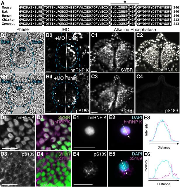
Figure 2.
Serine 189, the JNK target site on Xenopus hnRNP K, is phosphorylated in neurons in vivo. A, Multiple sequence alignment of the region of hnRNP K surrounding X. laevis hnRNP K S189 (indicated by *) from five species shows that the prospective JNK site is highly conserved. Black and gray shading indicates 100 and 80% conservation among the different species examined, respectively. The black line indicates the region of Xenopus hnRNP K used to generate the phospho-specific antibody (pS189). B, C, Immunohistochemistry (IHC) on transverse sections of spinal cord and somites of stage 37/38 embryos. B, Sections of embryos unilaterally injected with hnRNP K MO at the two-cell stage. Overlays indicate locations of spinal cord (SC) and somites (Som). Unilateral MO suppression of endogenous hnRNP K caused a similar reduction of immunostaining (p = 0.9, two-sided t test, 5 sections per embryo, n = 3 embryos per condition) for both total hnRNP K (55 ± 1% injected/uninjected, p = 0.03, one-sided t test as before; B2) and pS189 (56 ± 5% injected/uninjected, p = 0.01, one-sided t test as before; B4) on the injected side, indicating that pS189 is specific for hnRNP K. Phase contrast images (B1, B3) demonstrated that loss of staining in spinal cord and somites was attributable to loss of expression on the injected side and not to loss of cells. C, Sections of uninjected embryos treated with alkaline phosphatase before immunostaining for total hnRNP K (C2) or pS189 (C4), demonstrating specificity of pS189 for phosphorylated hnRNP K. C1, C3, SYBR Green staining of nuclei demonstrated that loss of staining was not attributable to cell loss. D, E, Representative images of immunostaining (magenta) of stage 37/38 hindbrain sections (D; confocal microscopy) and neurons from dissociated neural tube/myotome cultures (E; conventional microscopy) with the pS189 antibody indicated hnRNP K was phosphorylated at S189 in vivo during nervous system development. Whereas total hnRNP K shuttles to the cytoplasm but localized predominantly to the nucleus during axon outgrowth (D1, D2, SYBR Green; E1–E3, DAPI, blue), phospho-hnRNP K (pS189) was primarily cytoplasmic and perinuclear (D3, D4, E4–E6). Arrows indicate regions measured for intensity profiles (E3, E6). Scale bars, 20 μm.

