Figure 8.
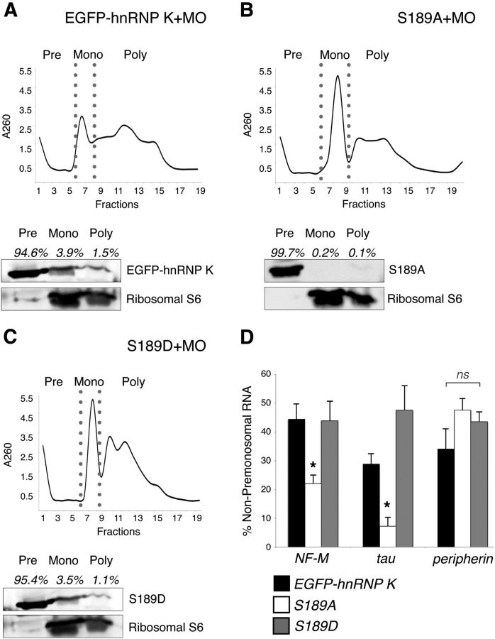
Phosphorylation of the JNK site on EGFP–hnRNP K regulates handoff of its RNA targets to the translational machinery. Polysome profiling followed by Western blot analysis of cytosolic extracts from embryos unilaterally coinjected with hnRNP K MO and either EGFP–hnRNP K (A), S189A (B), or S189D mRNA (C). The black line indicates RNA absorbance at 260 nm (A260) across fractions, and premonosomal (Pre), monosomal (Mono), and polysomal (Poly) fractions are indicated (top). Western blots were probed with anti-GFP to visualize the EGFP fusions and anti-S6 to demonstrate proper polysomal separation (bottom). A, EGFP–hnRNP K was most abundant in the premonosomal fractions (94.6%) but was also present to a lesser extent in the monosomal (3.9%) and polysomal fractions (1.5%). B, The loss of phosphorylation at the JNK site prevented S189A from moving beyond the premonosomal fraction (99.7%) into heavier, translating fractions (0.3%). C, S189D, like EGFP–hnRNP K (A), also appeared across all fractions (premonosome, 95.4%; monosome, 3.5%; polysome, 1.1%). D, Quantitation by real-time qRT-PCR of the target (NF-M and tau) and nontarget (peripherin) RNAs of hnRNP K among non-premonosomal fractions (monosome + polysome) with real-time qRT-PCR. Relative amounts (corrected against uninjected WT; see Results) of NF-M (*p = 0.04, one-way ANOVA with Tukey's post hoc test; 3 replicates, 25 embryos per sample) and tau (*p = 0.01, as before) mRNAs in these fractions for embryos unilaterally coinjected with hnRNP K MO and S189A RNA were significantly reduced compared with those coinjected with EGFP–hnRNP K or S189D RNA, whereas relative amounts of peripherin mRNA were not significantly different among groups (p = 0.2, one-way ANOVA as above). Error bars indicate pooled SDs. ns, Nonsignificant.

