
Figure 2.
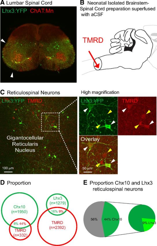
Lhx3 and Chx10 medRF neurons are reticulospinal neurons. A, Adult Lhx3-YFP upper lumbar spinal cord section with ChAT-positive motoneurons. Arrows indicate Lhx3-YFP axons in both ventrolateral and ventral funiculi. B, Schematic showing the application of retrograde tracer TMRD at cervical level in neonatal brainstem-spinal cord preparations. C, TMRD back-labeled Lhx3-YFP medRF neurons at low (left) and high magnifications (left, dashed window). White arrowheads indicate TMRD back-labeled Lhx3-YFP neurons. Yellow arrowheads indicate Lhx3-YFP-negative TMRD-positive reticulospinal neurons. D, Proportions of fluorescent protein-TMRD cells over fluorescent protein (green) and TMRD (red) cells (n = 3 Lhx3-YFP and n = 3 Chx10::GFP neonatal preparations). E, Percentages of Chx10 and Lhx3 neurons among TMRD-positive reticulospinal neurons.