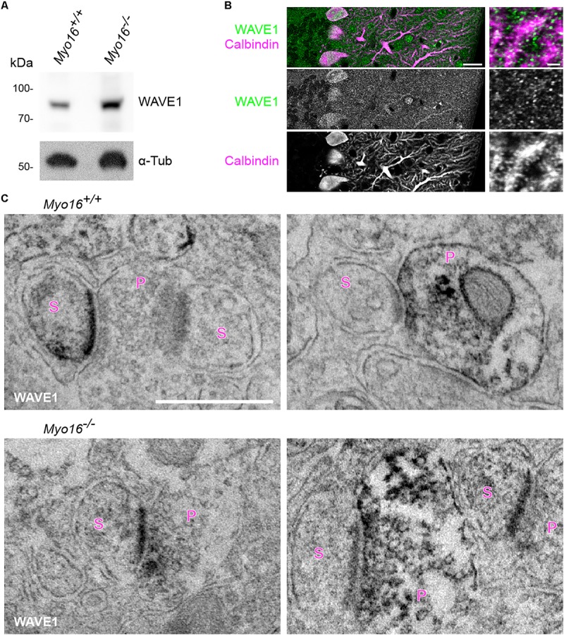
FIGURE 5.

WAVE1 localizes to Purkinje cell spines and to presynaptic terminals in the cerebellar molecular layer. (A) Western blot analysis of cerebellar extracts from 11 day old wild-type (Myo16+/+) and Myo16em3 knockout (Myo16–/–) mice. Antibodies against WAVE1 and α-Tubulin (α-Tub; loading control) were used. (B) WAVE1 partially co-localizes with PC spines. Confocal images of cerebellar sections from adult wild type mice immuno-fluorescently labeled with antibodies against WAVE1 and Calbindin-D-28K. Right panels are zoom-in images depicting the molecular layer. Scale bars, 20 μm (right panels); 2 μm (left panels). (C) Immuno-electron microscopy of cerebellar sections from adult wild-type mice (Myo16+/+; upper panels) and Myo16em3 knockout mice (Myo16–/–; lower panels) using WAVE1 antibody and DAB labeling. Black precipitate indicates presence of WAVE1. Left panels depict examples of PC spines positive for WAVE1, right panels are examples of presynaptic terminals positive for WAVE1. S, spine; P, presynaptic bouton. Scale bar, 500 nm.
