
Figure 2.
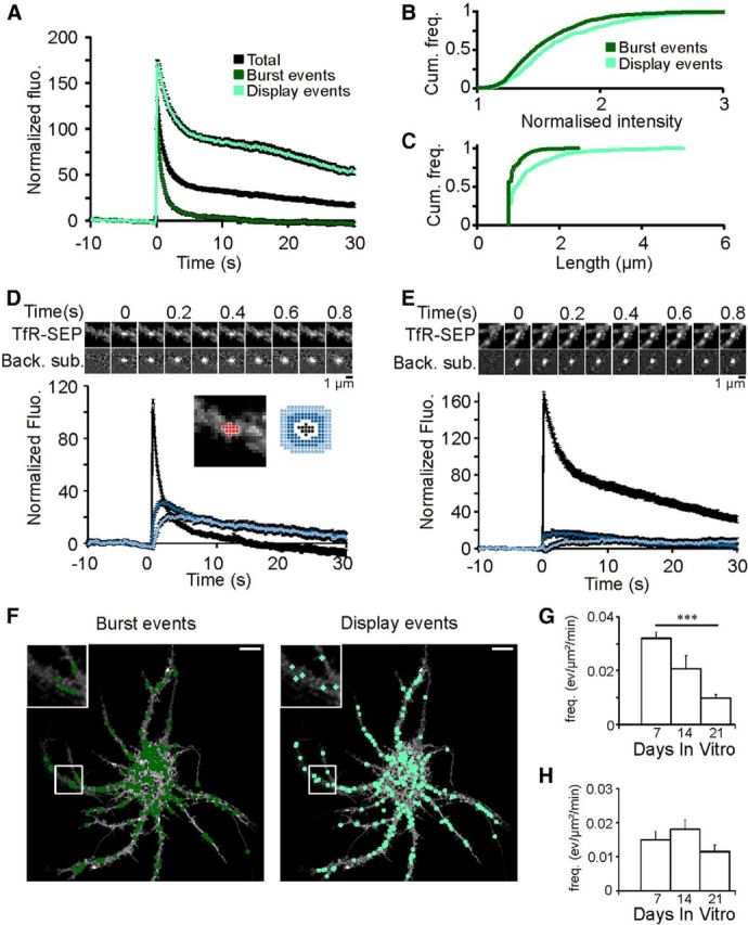
Properties of burst and display events in neuronal dendrites. A, Average of exocytosis events (black curve) recorded in 14–15 DIV neurons (n = 8), sorted in burst (dark green, 1139 events) and display (light green, 621 events). B, C, Maximal fluorescence (B) and length (C) of burst and display events. D, E, Diffusion of receptors after exocytosis. Total fluorescence measured in ROI (black), SR1 (dark blue), and SR3 (light blue) (see Materials and Methods), depicted in the scheme (inset), for burst events (D) and display events (E). Top, Consecutive images of example events. Raw images on top, background subtracted below. Scale bar, 1 μm. F, Map of the burst (n = 226) and display (n = 218) events detected on a neuron (14 DIV) during a 5 min movie at 1 Hz. Scale bar, 10 μm. G, H, Frequency of burst (G) and display (H) events recorded with 1 Hz imaging. Burst event frequency decreases with age in culture (significant difference between 7 and 21 DIV neurons, ***p < 0.001) but not display event frequency (H).