
Figure 2.
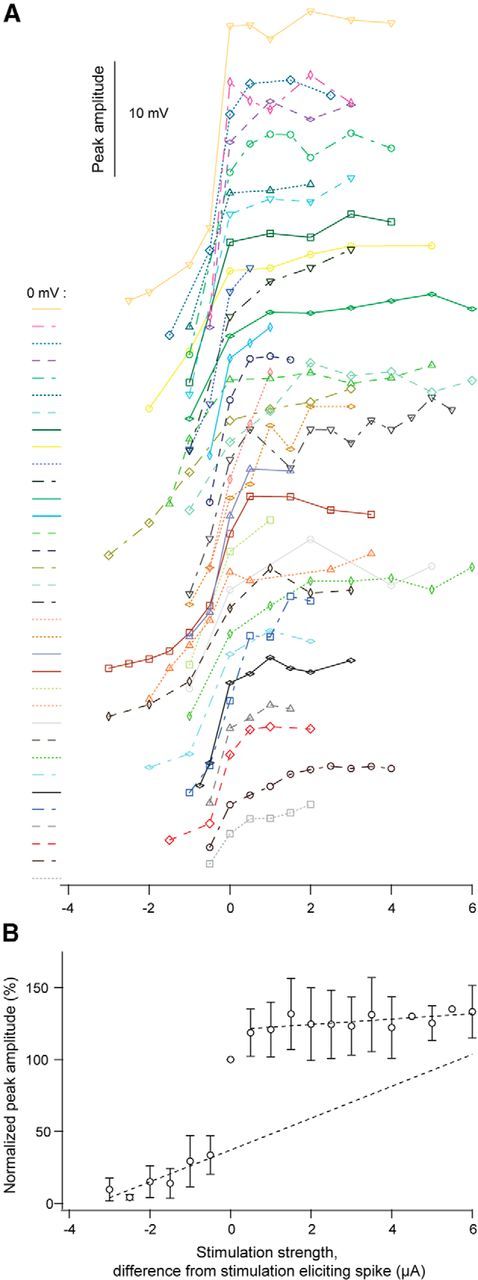
Changes of the amplitude of the potential by increasing strength of glutamate stimulation. A, Individual curves showing the peak amplitude with increasing strength of glutamate stimulation for all neurons. To the left of each curve the baseline (0 mV) is indicated together with a curve identifier (color and line type) for the specific curve. B, Normalized response amplitudes plotted against the glutamate stimulation strength for all neurons (mean ± SD; n = 34). Dotted lines: linear regression line fitted to the mean values below the spike threshold and to the mean values above the spike threshold.