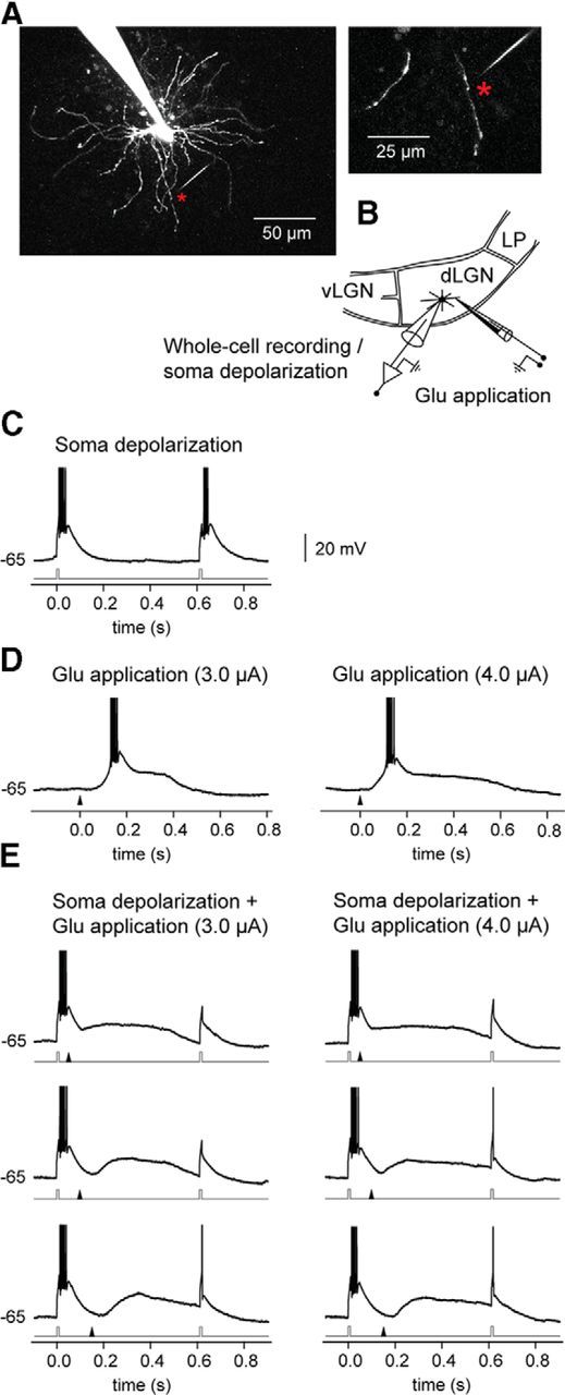
Figure 9.

A spike/plateau potential elicited at a single distal dendrite can inactivate T-type Ca2+ conductance in TC neurons thereby preventing burst firing. A, Image of the recorded TC neuron. The glutamate application site (indicated by an asterisk) was 67 μm from the soma and 5 μm from the dendrite. B, Scheme of experiment: simultaneous two-photon imaging and somatic whole-cell recording. vLGN, ventral lateral geniculate nucleus; LP, lateral posterior nucleus. C, T-type Ca2+ bursts were elicited by two depolarizing pulses (each 10 ms, 150 pA) to the soma of a hyperpolarized (−65 mV) TC neuron. The timing of the pulses is indicated by the current trace (gray) below the trace of the response. D, Responses, elicited by local dendritic glutamate stimulation (left, 3 μA; right, 4 μA). The time of the application of the glutamate pulse is indicated by the arrowhead below each trace. E, Responses elicited by combined somatic depolarizing pulses and dendritic glutamate stimulation. Due to the NMDA spike/plateau, a T-type calcium burst in response to the second somatic pulse was not elicited. The glutamate stimulation started 50 ms (top), 100 ms (middle), or 150 ms (bottom) after the first depolarizing pulse. Action potentials are truncated at 0 mV.
