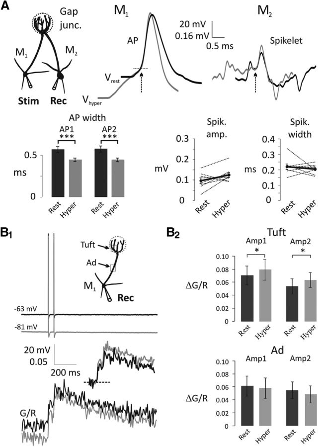
Figure 3.
Electrically coupled spikelets and bAP-evoked Ca2+ transients in the apical tuft of mitral cells are not decreased with somatic hyperpolarization. A, Somatic APs recorded from the presynaptic mitral cell (M1) narrow at Vhyper (representative average traces on the left; black, Vrest; gray, Vhyper). AP traces are overlaid by the AP generation threshold (dashed line). M2, postsynaptically recorded spikelets were aligned with the peak of the presynaptic AP and overlaid by the spikelet generation threshold (representative average traces on the right; black, Vrest; gray, Vhyper). Arrows indicate AP and spikelet initiation. Summary charts show AP width at half amplitude (bottom left; n = 8) and spikelet amplitude and width at half amplitude (bottom right). Black dots connected with thick black lines represent the means of eight individual experiments. ***p < 0.001. B1, Representative traces of a pair of bAPs (top) and Ca2+ transients (20 Hz, bottom) were measured in the apical tuft at Vrest (black) and Vhyper (gray). Sets of five sweeps at Vrest were alternated with sets of five sweeps at Vhyper. Inset, bAP-evoked Ca2+ transients are adjusted according to the basal [Ca2+]. B2, Summary chart shows Ca2+ transient amplitudes (Amp1, Amp2) at Vrest and Vhyper in the tuft and apical dendrite (Ad). Ca2+ transient amplitudes in the tuft (n = 7) are increased with hyperpolarization, although there is no significant change in the apical dendrite (n = 5). *p < 0.05.

