Figure 5.

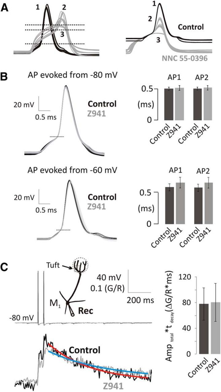
bAP-evoked Ca2+ transients are independent of T-type Ca2+ channels in mitral cells. A, NNC 55–0396 (50 μm) raises threshold (left; horizontal lines; 1, pre-NNC; 2, 9 min NNC; 3, 22 min NNC) and attenuates AP amplitude (right; overlaid at the AP generation threshold). B, Representative overlaid traces of somatic APs (threshold at horizontal line) and summary data indicate that the somatic AP shape is unchanged by Z941. Column charts represent the width at half-the threshold to peak amplitude of somatic APs in control (black) and in the presence of Z941 (10 μm; gray) delivered either at −80 (top; n = 5) or −60 mV (bottom; n = 5). C, bAPs (20 Hz, top) were delivered to mitral cells at Vhyper, and Ca2+ transients were recorded in the apical tuft in control (black) and in the presence of Z941 (gray). See location in the schematic drawing. Fitted single exponential curves indicate τdecay. Integrals of Ca2+ transients were measured as Amptotal × τdecay (n = 5, 5–20 min application).
