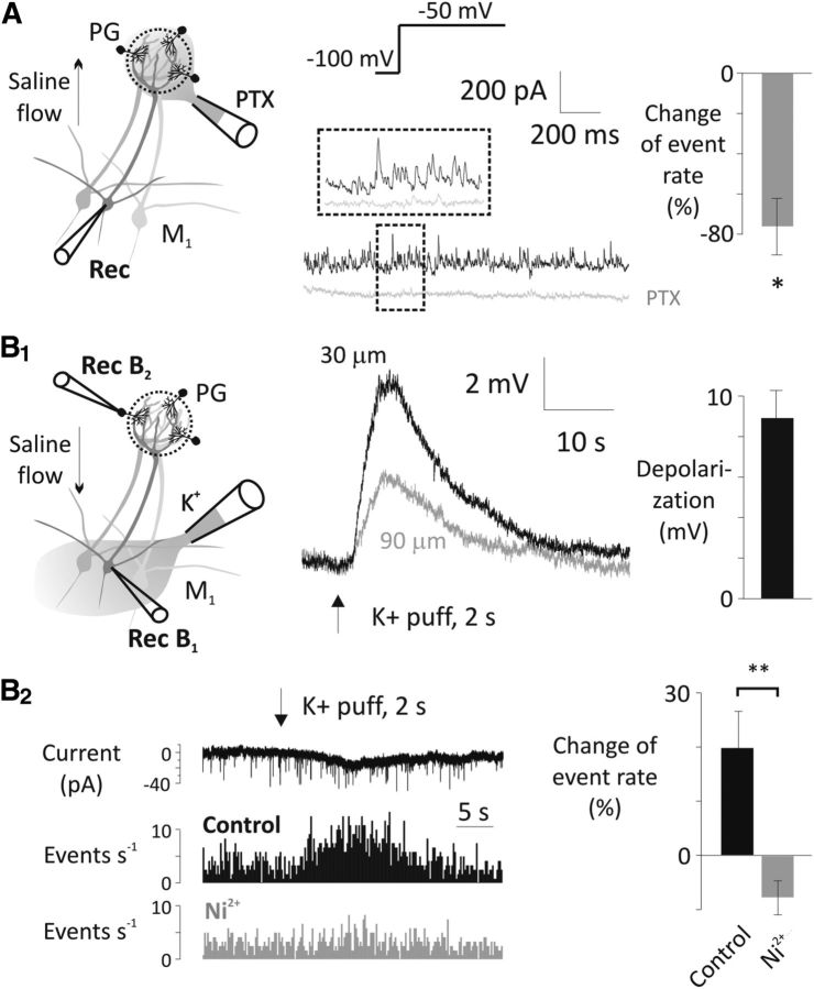
Figure 9.
Periglomerular cells are major sources of T-type Ca2+ channel-sensitive asynchronous GABA transmission onto mitral cells. A, Puff application of 50 μm PTX on the apical tuft of a recorded mitral cell (M1) reduced the frequency of IPSCs at −50 mV test potential after a 4 s hyperpolarizing prestep to −100 mV (n = 4). QX-314 (0.5 mm) was included in the pipette. PTX was directed away from the external plexiform layer with a laminar perfusion flow (see arrow on the drawing). Puffer pipette was positioned on the surface of the glomerulus. *p < 0.05. B1, A 2 s puff of 65 mm KCl solution from above the mitral cell layer (30 or 90 μm) depolarizes mitral cells (Rec B1, flow was directed away from external plexiform layer). B2, The high K+ solution puffed onto the mitral cell layer increased the EPSC rate in periglomerular cells (PG, Rec B2) as shown in the example current trace and the mini rate histogram (top and middle). This increase was abolished by 500 μm Ni2+ (bottom; n = 6, 6 trials before and after Ni2+ application in each cell). Again, high K+ solution was directed away from the glomerular layer with a laminar perfusion flow (see arrow on the drawing). TTX (1 μm), MK-801 (20 μm), and bicuculline (20 μm) were present throughout the mini rate measurements. **p < 0.01.

