Figure 1.
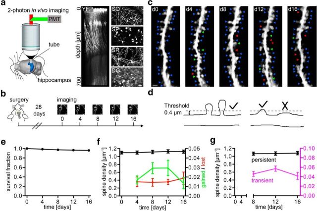
Structural plasticity of dendritic spines in the hippocampus. a, Left, Schematic illustrating two-photon in vivo imaging in the hippocampus. Right, Side projection of a z-stack acquired up to a depth of 700 μm below the hippocampal surface and maximum intensity projections from different depths in stratum oriens (SO), stratum pyramidale (SP), stratum radiatum (SR), and dentate gyrus (DG). b, Experimental timeline from surgery until imaging. c, Time-lapse images of a radial oblique dendrite over a period of 16 d. Note labeled stable spines (blue arrowheads), gained spines (green arrowheads), and lost spines (red arrowheads). d, Schematic with two examples for spine scoring procedure. e, Sixteen-day survival fraction of spines. f, Spine density of radial oblique dendrites in vivo (black) and densities of lost (red) and gained (green) spines. g, Density of transient and persistent spines. e–g: n = 3660 spines in 5 mice. Scale bars: a, 20 μm; c, 2 μm.

