Figure 6.
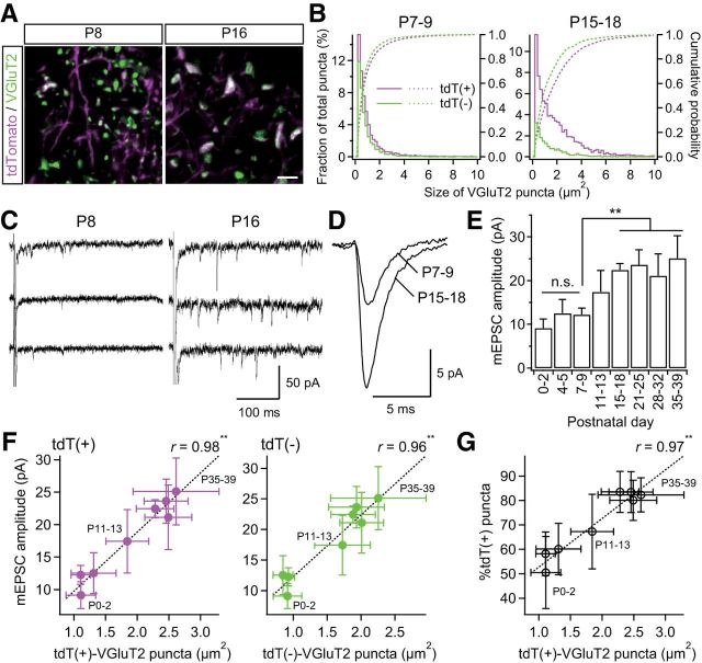
Strengthening of PrV2-origin lemniscal synapses during postnatal development. A, VGluT2-immunoreactive puncta in the V2 VPM before and after synapse elimination. Scale bar, 5 μm. B, Distributions and cumulative probabilities of tdT(+) and tdT(−) VGluT2 puncta before and after synapse elimination. Data from six and eight mice were pooled for P7–P9 and P15–P18, respectively. C, Miniature lemniscal EPSCs before and after synapse elimination. Miniature lemniscal EPSCs were evoked in the modified ACSF with Sr2+ ions instead of Ca2+ ions, whereas lemniscal fibers were stimulated electrically. D, Averaged miniature lemniscal EPSCs before and after synapse elimination. The numbers of averaged events were 489 and 763 for P7–P9 and P15–P18, respectively. E, The mean amplitude of miniature lemniscal EPSCs of a neuron significantly increased during the synapse elimination phase. Analysis was conducted on 512–814 events at 6–10 neurons from three mice for each developmental bin. F, The amplitude of miniature lemniscal EPSCs correlated well with the size of VGluT2 puncta during development. r, Pearson's correlation coefficient. Broken line is the regression line. G, Proportion of tdT-positive puncta correlated well with the size of tdT-positive puncta during development. Values are represented as mean ± SD. Statistical significance was tested by multiple t test with Bonferroni correction after one-way ANOVA for developmental changes in E and t test for noncorrelation for F and G. **p < 0.01; n.s., not significant, two-tailed test.

