Figure 3.
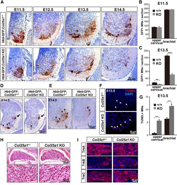
Motoneurons in Col25a1 KO mice underwent massive apoptosis during development. A, Anti-GFP immunostaining of the brachial spinal cords of Hb9-GFP;Col25a1+/+ (top) and Hb9-GFP;Col25a1 KO (bottom) embryos from E11.5 to E14.5. B, C, Quantitative analyses of GFP-positive motoneurons in the upper cervical (C3–C5) and brachial (C6–T1) spinal cord at E11.5 (B) and E13.5 (C). Data are mean ± SEM. ***p < 0.001. **p < 0.05. n = 3–5. D, Immunoperoxidase-labeled GFP-positive motoneurons (arrowheads) in the ventral horn and sympathetic preganglionic neurons (arrows) in the lateral horn of the thoracic spinal cord at E14.5. Representative images in multiple animals are shown. E, Immunoperoxidase-labeled GFP-positive motoneurons (arrowheads) in the ventral horn of the lumbosacral spinal cord at E14.5. Representative images in multiple animals are shown. F, TUNEL staining of the brachial spinal cord of Col25a1+/+ and Col25a1 KO embryos at E13.5. Arrowheads indicate TUNEL-positive apoptotic nuclei in the ventral horn. Nuclei were stained with DAPI (blue). G, Quantification of TUNEL-positive motoneurons in the brachial spinal cord at E13.5. Data are mean ± SEM. ***p < 0.001. n = 10. H, Overview (top) and high-magnification views (bottom) of H&E-stained sections of the DRG at E18.5. Representative images in multiple animals are shown. I, Immunostaining of DRG neurons with anti-TrkA, TrkB, and TrkC antibodies at E13.5. Nuclei were stained with DAPI (blue).

