
Figure 2.
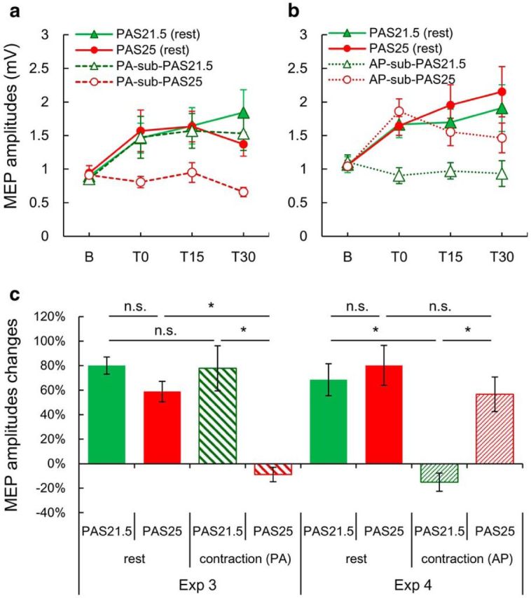
Comparison of the effects of standard PAS21.5 and PAS25 (PA pulses; suprathreshold TMS at rest) with subthreshold PAS21.5 and PAS25 evoked by PA- or AP-TMS pulses during voluntary contraction. a, b, Mean MEP amplitudes before (b) and after (T0, T15, and T30) different PAS protocols. a, Data from Experiment 3. MEPs were significantly increased compared with baseline at all time points by standard PAS21.5 (green filled triangle), PAS25 (red filled circle), and PA-sub-PAS21.5 (green triangle), but not PA-sub-PAS25 (red circle) (two-way rmANOVA: interaction PAS × STATE, F(1,8) = 22.518, p = 0.001; Tables 4 and 5). b, Data from Experiment 4. MEPs were significantly increased over baseline at all time points by standard PAS21.5 (green filled triangle), at T15 and T30 by PAS25 (red filled circle), and at T0 by AP-sub-PAS25 (red circle), but there was no MEP increase after AP-sub-PAS21.5 (green triangle) (two-way rmANOVA: interaction PAS × STATE, F(1,8) = 6.226, p = 0.037; Tables 4 and 5). c, Grand average (over T0, T15, and T30) MEP percentage changes over baseline MEPs in each session. Subthreshold PAS21.5 and PAS25 induced MEP facilitation through the repetitive activation of PA and AP inputs, respectively (four-way rmANOVA on the combined data from Experiments 3 and 4: interaction PAS × STATE × EXPERIMENT, F(1,64) = 9.044, p = 0.004; Table 4). *p < 0.05 (post hoc Turkey's tests). n.s., Not significant.