
Figure 3.
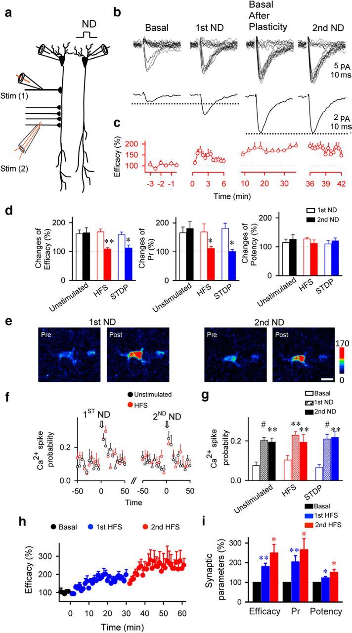
Astrocytic modulation of synaptic transmission is impaired after LTP protocol. a, Scheme of paired-recorded pyramidal neurons and the stimulating electrodes. b, Responses evoked by minimal stimulation (top traces) and average EPSCs (n = 60 stimuli, including successes and failures; bottom traces) before (basal), after first ND, 30 min after HFS (Basal After Plasticity), and after second ND. c, Synaptic efficacy (i.e., mean amplitude of responses including successes and failures of neurotransmission) before, after first ND, 30 min after HFS, and after second ND (n = 7). First and second NDs were delivered at 0 and 36 min, respectively. d, Relative changes from control basal values of synaptic parameters after first and second ND in unstimulated (n = 7), HFS (n = 7), and STDP (n = 5)-stimulated slices. Pr, Probability of release. e, Fluorescence intensities of Fluo-4-filled astrocytes before and after first and second ND. Scale bar, 10 μm. f, Astrocyte Ca2+ spike probability in unstimulated (111 astrocytes, n = 11 slices) and HFS conditions (71 astrocytes, n = 8 slices). Zero time corresponds to the beginning of ND. g, Ca2+ spike probability before (basal; white bars) and after first (striped bars) and second ND (n = 5; filled bars) in unstimulated (n = 11) and HFS (n = 8) or STDP (n = 6)-stimulated. h, Relative synaptic efficacy evoked by minimal stimulation versus time before (basal, black circles), after first stimuli (first HFS, blue circles), and after second stimuli (second HFS, red circles; n = 4). Zero time corresponds to the onset of first HFS. Note that the system is not saturated after first stimulation. i, Relative changes from control values (basal, black bars) of synaptic parameters after first (blue bars) and second (red bars) HFS (n = 4). Significant differences were established at *p < 0.05, **p < 0.01, and #p < 0.001.