Figure 10.
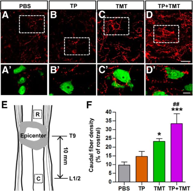
Density of serotonergic (5-HT) axons in the caudal lumbar motor region. A–D, Serotonergic (5-HT) axons in the lumbar ventral motor areas were visualized by immunofluorescence staining in longitudinal sections of the injured spinal cord at 9 weeks after injury. After SCI, the density of 5-HT axons in the region caudal to the lesion was markedly reduced (A). Animals in the TP + TMT group showed the most robust increase in 5-HT axon density (D). A′–D′, Magnified images of the boxed regions in A–D. Neurons were visualized by NeuN staining (green). Spinal motor neurons were frequently contacted by 5-HT axon terminals in TMT-alone and TP + TMT groups. Scale bars, 50 and 20 μm for magnified images. E, Illustration of the location of regions of interest (boxed) in the longitudinal plane. The center of the caudal motor regions was, on average, 10 mm apart from the epicenter. Rostral regions of interest were used to control for differences in the intensity of immunoreactive signals between individual sections. F, Quantification of the caudal 5-HT axon density as a percentage of that in the rostral region. *p < 0.05 and ***p < 0.01 compared with PBS group; ##p < 0.01 compared with the TP-alone group by one-way ANOVA followed by Tukey's post hoc analysis. n = 8 animals per group.

