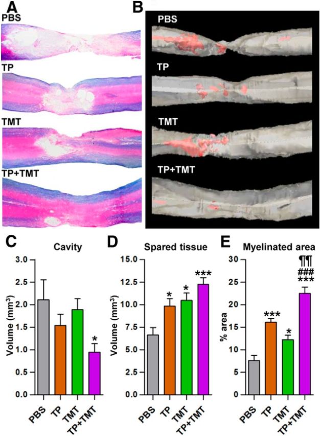
Figure 9.

Structural repair of injured spinal cord at the lesion level by combined NSC transplantation and TMT. A, Representative images of the lesion epicenter region stained with eriochrome cyanine RC to visualize the spared white matter and eosin counter-staining in the injury-alone (PBS), TP-alone, TMT-alone, and TP + TMT groups. Animals were killed 8 weeks after injury. B, 3D reconstruction images from the same animals as in A. Red color represents cystic cavity. C–E, Comparison of the lesion cavity volume (C), spared tissue volume (D), and percentage of myelinated area (E) within the spared spinal cord tissue. *p < 0.05 and *p < 0.001 compared with the injury-alone group (PBS), ¶¶p < 0.01 compared with the TP-alone group, and ###p < 0.001 compared with the TMT-alone group, by one-way ANOVA followed by Tukey's post hoc analysis. n = 8 animals per group for all analyses.
