Figure 3.
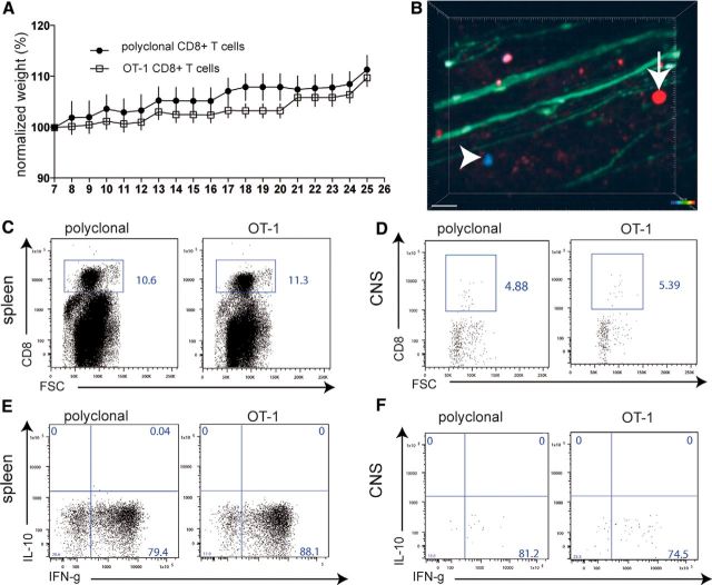
Transfer of activated OT-1 CD8+ T cells into lymphopenic mice. OT-1 spleen cells were activated by 50 nm OVA257–264 in the presence of IL-12 and IL-18. As the control, anti-CD3/anti-CD8 polyclonally activated wild-type C57BL/6 spleen-derived CD8+ T cells were similarly activated. A total of 5 × 10⁶ activated CD8+ T cells were injected on day 7 of culture into naive lymphopenic Rag1−/− mice (n = 8, data representative of two experiments). A, CD8+ T-cell-transferred Rag1−/− mice were monitored for weight loss for 26 d and clinically evaluated according to EAE function scores. B, TPLSM of the brainstem of living anesthetized Rag1−/−/thy1-EGFP mice, which had been injected with red-fluorescent OT-1 cells and blue fluorescent pcCD8+ T cells, showed only a few, most likely perivascular, activated OT-1 T cells and control pcCD8+ T cells in the CNS without signs of CNS tissue invasion (representative of six imaging areas from two experiments; scale bar, 30 μm). C, On day 25, immune cells were isolated from spleens in which large numbers of the transferred CD8+ T cells could be found. D, In the same animals as in C, only very few cells in total and low numbers of transferred CD8+ T cells (OT-1 or pcCD8+ T cells) could be found by flow cytometry in CNS isolated immune cells (single dot represent single cells; pooled CNS samples from three animals). E, Upon restimulation with plate-bound anti-CD3/anti-CD28 antibodies, strong IFN-γ production was recalled in the same way for both control and OT-1 CD8+ T cells isolated from the spleen or the CNS (F).

