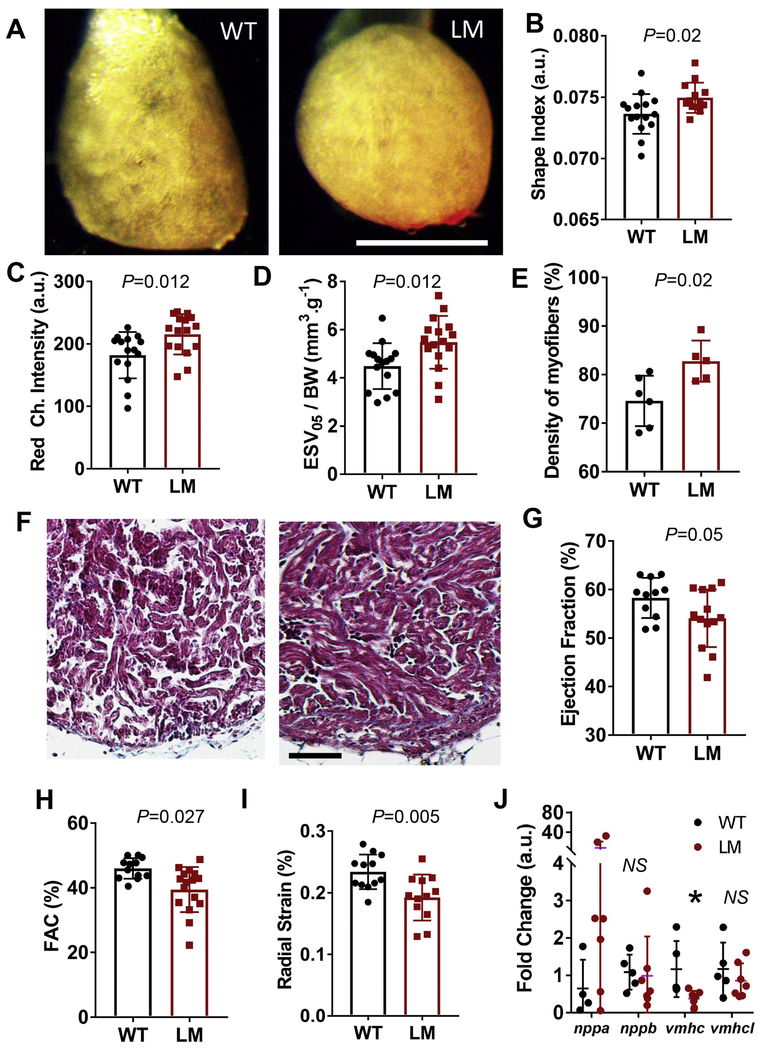
Fig. 3. Ventricular remodeling in lamp2e2/e2 hearts.
A. Images of isolated ex vivo perfused hearts. Ventricles in lamp2e2/e2 appear rounder. Scale bar is 1 mm. B. Shape index (area over perimeter squared) is increased in lamp2e2/e2 fish (N=15 for wild-type and 16 for lamp2e2/e2). C. Red channel intensity is bigger in lamp2e2/e2 hearts, supporting denser tissue (N=15 for wild-type and 16 for lamp2e2/e2). D. Increased end-systolic volume at low flow (0.05 ml/min; ESV05) in lamp2e2/e2hearts (N=15 for wild-tvpe and 16 for lamp2e2/e2). E. Quantification of F (N=6 hearts for wild-type and N=5 for lamp2e2/e2). F. Trichrome-stained heart slices show denser trabeculae myocardium. Scale bar is 50 μm. G. Reduced ejection fraction (EF%), H. Reduced fractional area contractility (FAC%), and I. reduced radial strain both suggest compromised cardiac pump function in lamp2e2/e2 (LM). Shown in G-I are ex vivo studies of Langendorff-like perfused hearts (N=12). J. qRT-PCR to quantify transcripts of fetal gene program (N=5 and 7 for WT and LM, respectively). *P<0.05.

