
Figure 2.
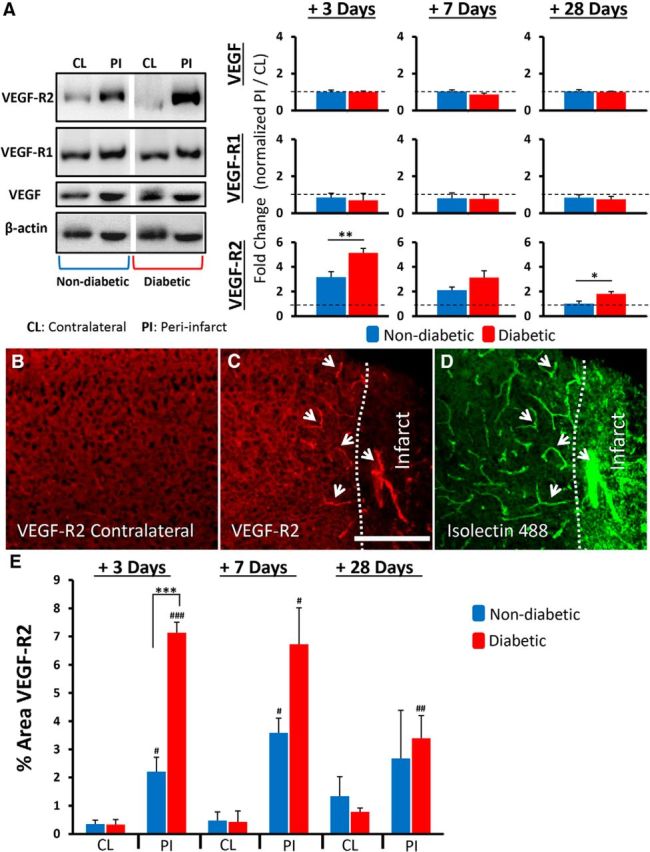
Diabetes amplifies the upregulation of VEGF-R2 after stroke. A, Representative Western blot (cropped) from tissue 3 d after stroke and quantification of VEGF and its receptors 3, 7, and 28 d after stroke in peri-infarct (PI) cortex (normalized to β-actin) relative to undamaged contralateral (CL) hemisphere (normalized to β-actin) (n = 5–7 mice per group). Immunohistochemical staining for VEGF-R2 in the contralateral hemisphere (B) and peri-infarct cortex (C). VEGF-R2 expression colocalizes with isolectin B4-labeled vessels in the peri-infarct cortex (D, see white arrows). Scale bar, 200 μm. E, Quantification of VEGF-R2 expression in peri-infarct or contralateral cortex (n = 4–6 mice per group). *p < 0.05, comparing nondiabetics with diabetics. **p < 0.01, comparing nondiabetics with diabetics. ***p < 0.0001, comparing nondiabetics with diabetics. #p < 0.05, comparing contralateral with peri-infarct.##p < 0.01, comparing contralateral with peri-infarct.###p < 0.001, comparing contralateral with peri-infarct.