
Fig. 4.
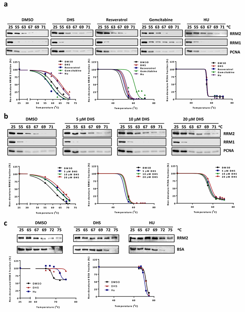
DHS interacts with RRM2. a Cellular thermal shift assay to examine interactions of compounds (10 μM DHS, 10 μM resveratrol, 0.5 μM gemcitabine, or 500 μM HU) with RRM1 and RRM2. Cells were treated with 10 μM MG132 for 1 hour followed by 4-hour incubation with compounds before performing thermal shit assay. Low panel is the charts of percentages of non-denatured protein fraction. b Cellular thermal shift assay to analyze the interaction of DHS (5, 10, or 20 μM) with RRM2. Low panel is the charts of percentages of non-denatured protein fraction. c In vitro thermal shift assay to analyze DHS (10 μM) and RRM2 interaction in vitro. DHS or HU (500 μM) and 400 ng purified RRM2 were incubated for 4 hours. The aliquots were further incubated at 65, 67, 69, 72, or 75 °C for 4 minutes. Low panel is the charts of percentages of non-denatured protein fraction.