
Figure 1.
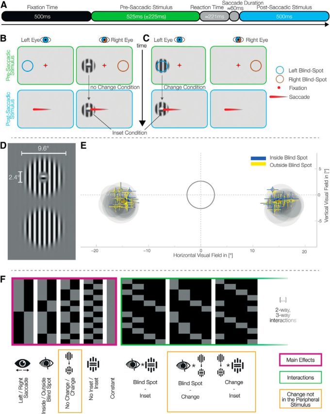
Experimental setup and eye-movements behavior. A, Trial time course. B, C, Each set of two panels represents what is presented to each eye with the shutter glasses. After a fixation interval, a stimulus appeared monocularly in the periphery (top). After the disappearance of the fixated crosshair, the subjects perform a saccade to the center of the presaccadic stimulus, which becomes the postsaccadic stimulus (bottom). The colored circles represent the location of the blind spot in each eye and were not displayed on the screen. B, An example of a trial without change: the inset stimulus, presented outside the blind spot, does not change before and after the saccade. Importantly, presenting an inset stimulus inside the blind spot always leads to fill-in and therefore the perception of a continuous stimulus. We therefore recorded the inset no-change condition only outside the blind spot. C, A trial with change: the continuous stimulus, presented inside the blind spot, is exchanged during the saccade to an inset stimulus. D, Gabor patches used as stimuli; horizontal stimuli were also used. The inset was set to ∼50% the diameter of the blind spot. E, Locations of the blind spots and saccades' end-points. The gray ring encloses the tolerance area for fixation. The gray discs represent the average calibrated blind spot sizes and locations for each subject. Bold crosses represent the winsorized average saccade end locations over subjects of both inside and outside blind spot trials (±winsorized SD). Small crosses show the same metrics for each individual subject. F, The design matrix used for the single subject GLMs. An overparameterized model of four main effects (purple), constant, and all interactions (green) was used.