
Figure 2.
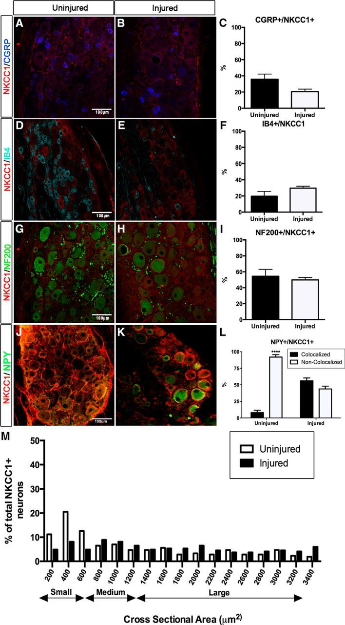
Immunohistochemical analyses of NKCC1 expression in DRG neuron subpopulations in injured and uninjured animals. A, B, D, E, G, H, J, K, Confocal images of unmyelinated peptidergic (CGRP+; A, B), nonpeptidergic (IB4+; D, E), myelinated (NF200+; G, H), and NPY+ (J, K) DRG neurons expressing NKCC1 in injured and uninjured rats. Scale bar, 100 μm. C, F, I, L, There were no significant differences in the quantification of NKCC1 expression between injured and uninjured animals (C, F, I), except for the coexpression of NPY (L). Data are presented as the mean ± SEM. J, Morphometric analysis of NKCC1+ neurons in uninjured (white) and injured (black) DRGs. M, The frequency distribution of soma diameter of neurons expressing NKCC1 shows no differences between uninjured and injured animals.