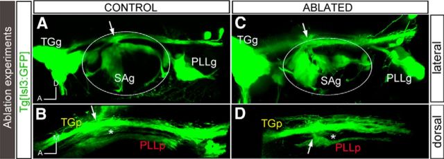
Figure 4.
Ablation of the first differentiated neurons results in defects in the sensory projections at the central level. Tg[Isl3:GFP] embryos were used for the ablation of the first differentiated neurons from the ALLg/SAg using the laser of a multiphoton microscope. A, C, Lateral views of control and ablated embryos, respectively, showing no ectopic entry points after pioneer axon ablation. B, D, Dorsal views of A and C, respectively, showing the sensory projections at the central levels. Note the defects in SAg nerve bundle elongation upon ablation (compare B, D, asterisks). White arrows indicate the entry point. White asterisks indicate the location of the SAp. The contour of the otic vesicle is indicated with white circles. Anterior is always to the left.

