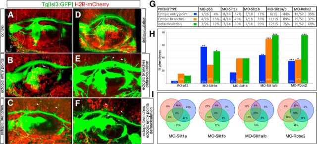
Figure 8.
Robo2/Slit1 signaling regulates axonal branching and nerve bundle fasciculation. Tg[Isl3:GFP] embryos were coinjected with MO-CTRL (MO-p53), mRNA for H2B-mCherry or lyn-TdTomato, and MO-Slit1a, MO-Slit1b, MO-Robo2, or double MO-Slit1a/b. A–F, Examples of phenotypes observed at 48 hpf. Note the variety of effects ranging from ectopic entry points (white arrows), ectopic branches (white arrow heads), defasciculation (black asterisks), and combinations of primary phenotypes (E, F). G, Statistics of MO injections. H, Analyses of the percentages of different phenotypes with different MO combinations. *p < 0.1; **p < 0.01; ***p < 0.001. I, Analysis of the percentage of morphant embryos displaying different combinations of phenotypes. Orange circles correspond to embryos displaying ectopic branches, blue circles to embryos with ectopic entry points, and green circles to embryos with defasciculation. Note that many embryos display a combination of phenotypes.

