
Figure 4.
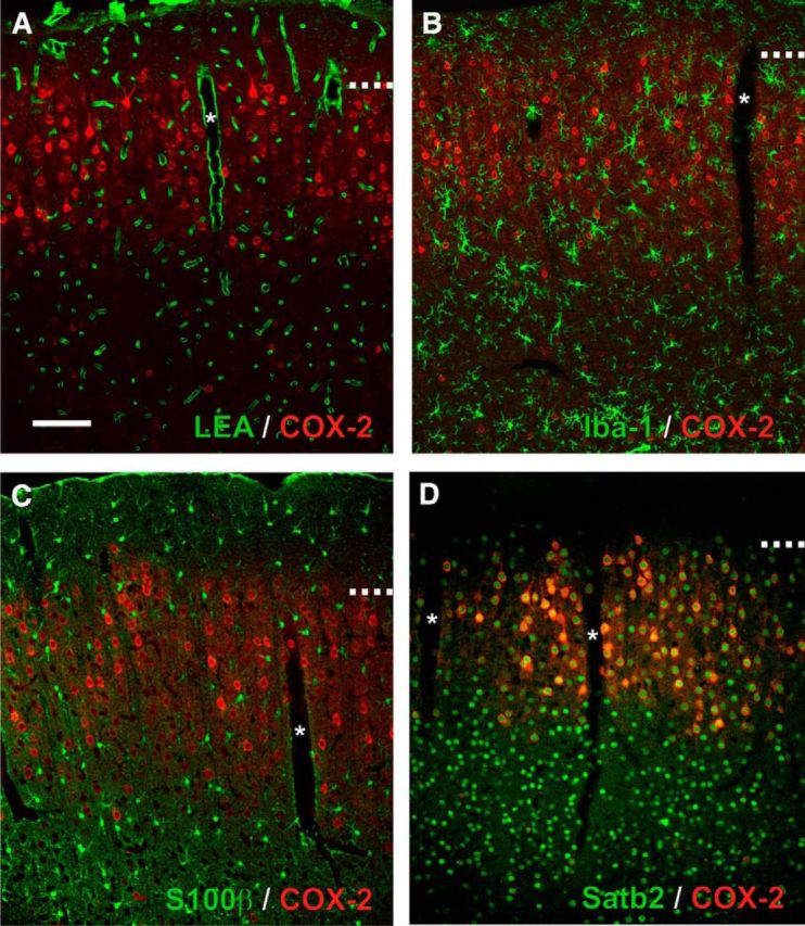
Expression of COX-2 in the neuro-glio-vascular unit. Representative single plane confocal images of double fluorescence staining showing the constitutive expression of COX-2 (red). Scale bar, 100 μm. *Denotes diving blood vessels. Dashed lines represent layer I–II border. Pial surface is upward. A, COX-2 immunolabeling is absent from the vascular bed stained with LEA (green) and (B) from microglia immunostained for Iba-1. C, S100β immunostained astrocytes (green) are essentially COX-2-negative. D, COX-2-immunopositive cells are Satb2-positive (green).