Figure 1.
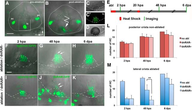
RAR activity is required for hair cell regeneration in the inner ear. A–D, Inner ear of 4.5-d-old tg(brn3c:mGFP) larvae with GFP labeling of hair cells in the three sensory cristae before (A) and after (B) lateral crista hair cell 2-photon laser ablation (detail, right). C, C′, High magnification view of the lateral crista showing hair cell bodies and kinocilia labeled with membrane-GFP. D, A transient air bubble over the crista is generated after laser ablation. E, Schematic representation of experimental procedure to assess the role of RA in hair cell regeneration in the inner ear. Briefly, after laser ablation of the lateral crista, two heat-shocks were performed to induce dnRAR expression, and hair cell number was quantified at 2 hpa, 48 hpa, and 6 dpa. F–K, Representative examples of hair cell regeneration in the lc after laser ablation in non-dnRAR (F–H) and dnRAR-expressing larvae (I–K). G, J, Arrows indicate kinocilia of the ablated crista at 48 hpa. J, K, dnRAR-GFP is visible in the inner ear. L, M, Quantitative analysis of the number of hair cells at different time points in nonablated posterior crista (L) and ablated lateral crista (M). A, Anterior; D, dorsal; ac, anterior crista; lc, lateral crista; pc, posterior crista. **p < 0.01. Scale bars: A, 50 μm; F, 10 μm.

