
Figure 4.
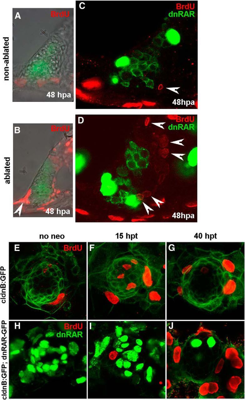
The RA pathway is required for supporting cell proliferation. A–D, To evaluate the role of RA in proliferation, lateral cristae of 4.5-d-old tg(brn3c:mGFP) and tg(brn3c:mGFP;hsp70:dnRARaa-GFP) larvae were laser-ablated and heat-shocked to induce dnRAR-GFP expression after 1 h. A, B, Increase of BrdU-positive cells was detected 48 hpa in treated larvae compared with the nonablated controls. C, D, Again, cell proliferation is induced in ablated lateral crista, and only non-dnRAR cells can enter the cell cycle (arrowheads). E–J, To confirm the results from the inner ear, 4.5-d-old tg(claudinb:GFP) and tg(claudinb:GFP;hsp70:dnRARaa-GFP) larvae were treated with neomycin and heat-shocked to induce dnRAR-GFP expression. E–G, The number of BrdU-positive cells (red) increased significantly 15 h after neomycin-induced hair cell damage in non-dnRAR larvae (F). At 40 hpt, cell proliferation was mainly detected in mantle cells (G). H–J, Cell proliferation is very low at 15 hpt in dnRAR-overexpressing larvae, and only non-dnRAR cells can enter the cell cycle (non-green cells) (I). At 40 hpt, less dnRAR is present and extensive proliferation is found in external cells (J).