Figure 6.
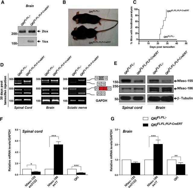
QKIFL/FL; PLP–CreERT mice display Nfasc pre-mRNA splicing defects. A, PCR amplification of genomic DNA from brains of 8-week-old wild-type and QKIFL/FL;PLP–CreERT mice injected with OHT for 5 consecutive days to confirm the Cre-mediated recombination and the presence of a 1 lox band. Mice were killed, and brains were collected at 30 d after OHT injection. B, Photographs of QKIFL/FL;− and QKIFL/FL;PLP–CreERT mice 30 d after OHT injection. C, Kaplan–Meier plot of onset of paralysis in QKIFL/FL;− (n = 11) and QKIFL/FL;PLP–CreERT (n = 11) mice. The statistical significance was assessed by log-rank (p < 0.0001). D, RT-PCR analysis of spinal cords, brains, and sciatic nerves from wild-type and QKIFL/FL;PLP–CreERT mice injected with OHT at 30 d after injection using primers flanking exon 17 (inclusion denotes Nfasc186) or exons 21/22 (inclusion denotes Nfasc155). E, Immunoblot analysis of lysates obtained from spinal cord and brain samples of 8-week-old wild-type and QKIFL/FL;PLP–CreERT mice injected with OHT for 5 consecutive days and harvested at 30 d after OHT injection. Blots were against anti-Nfasc155, anti-Nfasc186, and anti-β-tubulin. F, RT-qPCR analysis from spinal cord samples (n = 4) of 8-week-old wild-type and QKIFL/FL;PLP–CreERT mice injected with OHT for 5 consecutive days and collected at 30 d after OHT injection. Primers were specific for qkI, Nfasc155 ex21/22, and Nfasc186 ex17 mRNAs. The results are represented in terms of the mean of the fold change after normalizing the mRNA levels to GAPDH mRNA ± SEM. *p < 0.05; ***p < 0.001. G, RT-qPCR analysis from brain samples (n = 4) of 8-week-old wild-type and QKIFL/FL;PLP–CreERT mice injected with OHT for 5 consecutive days and collected 30 d after OHT injection. Primers were specific for qkI, Nfasc155 ex21/22, and Nfasc186 ex17 mRNAs. The results are represented in terms of the mean of the fold change after normalizing the mRNA levels to GAPDH mRNA ± SEM. **p < 0.01; ***p < 0.001.

