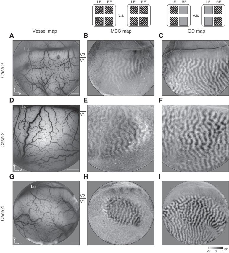
Figure 4.

MBC imaging cases. Three more cases imaged with MBC stimuli are shown. The three columns are surface blood vessel maps (left), MBC maps (middle) obtained by comparing MBC in left-eye (LE) conditions and MBC in right-eye (RE) conditions (similar to Fig. 3B), and OD maps (right) obtained by comparing left-eye and right-eye monocular stimulation conditions (similar to Fig. 1B). A–C, Images from Case 2, in which MBC stimuli was a 1.5° disk on a 5° background. D–F, Images from Case 3, in which MBC stimuli was also a 1.5° disk on a 5° background. In this case, the exposed V1 cortex was closer to the fovea region, and thus the same 1.5° MBC disk corresponded to a larger V1 map. G–I, Images from Case 4, in which MBC stimuli was a 1.5° disk on a 7° background. All three cases show a biased eye activation elicited by the MBC disk. No filtering was applied to these maps. Lu, Lunate sulcus. Scale bars: 2 mm.
