Figure 7.

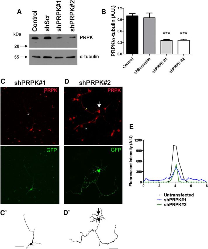
PRPK knockdown in neuroblastoma and primary neurons. A, N1E-115 cells were transfected with PRPK WT or were cotransfected with PRPK WT and either shScr, shPRPK#1, or shPRPK#2. Untransfected cells were included as an additional control. After 48 h, PRPK expression levels were assessed by immunoblotting using anti-PRPK and α-tubulin. B, Quantification of PRPK expression from A. n = 3; one-way ANOVA with Dunnett's post-test (***p < 0.001). C, D, Cultured hippocampal WT neurons at 2 DIV, expressing shPRPK#1 (C) or shPRPK#2 (D). Transfected neurons are shown in C′ and D′; a neuron in D′ is also indicated by an arrow. Scale bar, 50 μm. E, PRPK fluorescence intensity along the white lines in C and D (axonal sections of neurons expressing shPRPK#1 or #2) and yellow line (axonal sections of an untransfected neuron).
