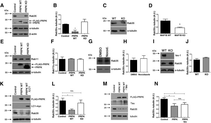
Figure 9.
PRPK regulates Rab35 expression levels. A, Rab35 expression levels evaluated by immunoblotting in COS7 cells transfected with PRPK WT or PRPK KD. Untransfected cells were used as a control. B, Quantification of Rab35 expression shown in A. n = 3; one-way ANOVA with Dunnett's post-test (*p < 0.05). C, Rab35 expression levels in embryonic brain from MAP1B WT or KO mice. D, Quantification of Rab35 expression shown in B. n = 3; unpaired Student's t test (*p < 0.05). E, Rab11 expression in COS7 cells expressing PRPK WT or PRPK KD. Untransfected cells were used as a control. F, Quantification of Rab11 expression shown in E. n = 3; one-way ANOVA with Dunnett's post-test (no statistically significant differences were observed). G, Two DIV cultured neurons were treated with DMSO or nocodazole 20 μm for 4 h, and then processed for immunoblotting. Rab35 expression remains unaffected between conditions. H, Quantification of Rab35 expression shown in C. n = 3; unpaired Student's t test (no statistically significant differences were observed). I, Brain protein extracts derived from WT and tau KO mice were analyzed with anti tau-1 and anti Rab35. In the KO brain, tau-1 immunoreactivity was absent, and Rab35 levels were unmodified. J, Quantification of Rab35 expression from A. n = 3; unpaired Student's t test (no statistically significant differences were observed). K, COS7 cells expressing PRPK WT alone or cotransfected with PRPK WT and LC1-myc were used to assess Rab35 expression levels. L, Quantification of Rab35 expression. n = 3; unpaired Student's t test (*p < 0.05). M, COS7 cells expressing PRPK WT alone or cotransfected with PRPK WT and Tau were used to assess Tab35 expression levels. N, Quantification of Rab35 expression. n = 3; unpaired Student's t test (*p < 0.05).

