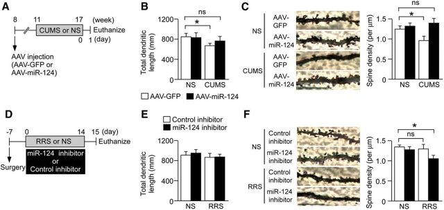
Figure 3.
miR-124 is associated with chronic stress-induced changes in dendritic morphology and spine density. A, Schematic of the experimental design used to assess the effects of miR-124 overexpression on stress-induced neuroplastic changes. Mice were injected with AAV–miR-124 or AAV–GFP into the bilateral hippocampus. Three weeks later, mice were subjected to a 6-week CUMS session. B, Bar graph showing the total dendrite length of DG granule neurons (n = 5–6 mice per group). CUMS reduced dendritic length in AAV–GFP-injected mice. Overexpression of miR-124 had no effect on dendritic length in NS mice but blocked the CUMS-induced decrease. *p < 0.05. C, Representative images of dendritic spines from DG granule neurons. Bar graph shows mean spine densities of DG granule neurons (n = 5–6 mice per group). CUMS reduced spine density compared with NS mice injected with the control vector, whereas miR-124 overexpression blocked this effect. *p < 0.05 versus NS. Scale bars, 10 μm. D, Schematic of the experimental design used to evaluate the effects of miR-124 inhibition on stress-induced neuroplastic changes. Mice were injected with miR-124 inhibitor or control inhibitor into the bilateral hippocampus every 3 d during 14 d of mild RRS (1 h/d). E, Bar graph showing the total dendrite length of DG granule neurons (n = 5–6 mice per group). This mild stress protocol had no effect on dendritic length. *p < 0.05. F, Representative images of dendritic spines from DG granule neurons. Bar graph shows spine densities of DG granule neurons after mild RRS with miR-124 inhibitor or control inhibitor cotreatment (n = 5–6 mice per group). The mild RRS regimen alone did not reduce spine density. Spine density was reduced in mild RRS-exposed mice injected with the miR-124 inhibitor, indicating enhanced susceptibility to stress-induced neuroplastic changes. *p < 0.05 versus NS. Scale bars, 10 μm. Data are presented as mean ± SEM.

