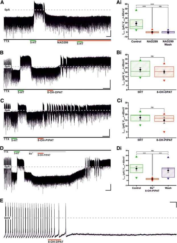
Figure 4.

Serotonin inhibits TIDA neurons via activation of the 5-HT 1A receptor. A, Voltage-clamp, oscillating TIDA neuron recorded as in Figure 3A. 5-HT is applied before, during, and after the 5-HT 1A antagonist, NAD299, is present in the bath. Note the complete abolishment of 5-HT response by NAD299. Calibration: 20 pA, 60 s. Ai, Box plot organized as Figure 3Bii, illustrating irreversible I5-HT blockade by NAD299 (n = 12). B, Voltage-clamp trace, oscillating TIDA neuron recorded as in Figure 3A. Sequential application of 5-HT and the 5-HT1A agonist, 8-OH-DPAT; note outward current induced by both compounds. Calibration: 20 pA, 60 s. Bi, 5-HT and 8-OH-DPAT induce statistically indistinguishable outward currents in TIDA neurons. Box plot organized as Figure 3Bii (n = 10). C, Voltage-clamp trace, oscillating TIDA neuron recorded as in Figure 3A. Sequential application of 5-HT and the 5-HT1A agonist, 8-OH-PIPAT; note outward current induced by both compounds. Calibration: 20 pA, 60 s. Ci, 5-HT and 8-OH-PIPAT induce statistically indistinguishable outward currents in TIDA neurons. Box plot organized as Figure 3Bii (n = 5). D, Voltage-clamp, oscillating TIDA neuron recorded as in Figure 3A (note abolishment of oscillation in response to TTX). Following a first exposure to 5-HT, eliciting a reversible outward current, Ba+ (300 μm) is bath applied to the cell. Note the absence of a response to 8-OH PIPAT (compare C) in the presence of Ba+ and how, as a consequence of the enduring nature of its interaction with the 5HT-1A receptor, the drug response is fully uncovered following wash of Ba+ from the recording chamber. Calibration: 20 pA, 60 s. Di, Ba+ significantly and reversibly reduces I8-OH-PIPAT. Box plot organized as Figure 3Bii (n = 9). E, Current-clamp, application of 8-OH-PIPAT induces cessation of oscillation and AP discharge. There is lack of recovery following wash of 8-OH-PIPAT. Calibration 20 mV 60 s. ***p < 0.005. ns, Not significant.
