Figure 6.

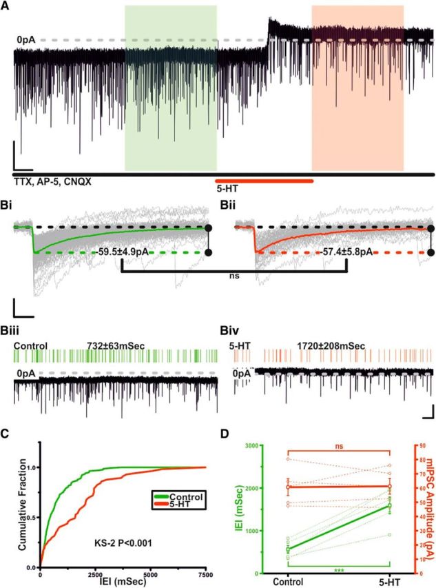
Serotonin inhibits mIPSCs in TIDA neurons via a presynaptic mechanism. A, Voltage-clamp trace showing mIPSCs from an oscillating TIDA neuron, isolated as in Fig. 5A, but with TTX added to abolish AP discharge. Application of 5-HT reversibly induces an outward current and a decrease in mIPSC frequency. Calibration: 40 pA, 20 s. Bi, Bii, Rise aligned mIPSCs (gray) with averaged event superimposed from boxed areas in A. Green represents Control. Red represents 5-HT. 5-HT application fails to alter mean mIPSC amplitude. Calibration: 30 pA, 10 ms. Biii, Biv, Enlarged views of the 100 s sections boxed in A with corresponding raster plots above. Green represents Control. Red represents 5-HT. Calibration: 40 pA, 5 s. C, Cumulative probability curves of mIPSCIEIs generated from the control (green) and 5-HT (red) sections boxed in A. The distribution shifts to significantly greater IEIs (K-S2; p < 0.001). D, 5-HT reversibly and significantly increases mIPSC IEI (green), whereas amplitude remains unchanged (red). Thick lines indicate mean ± SEM. Dashed lines indicate raw data (n = 5). ns, Not significant.
