Figure 4.
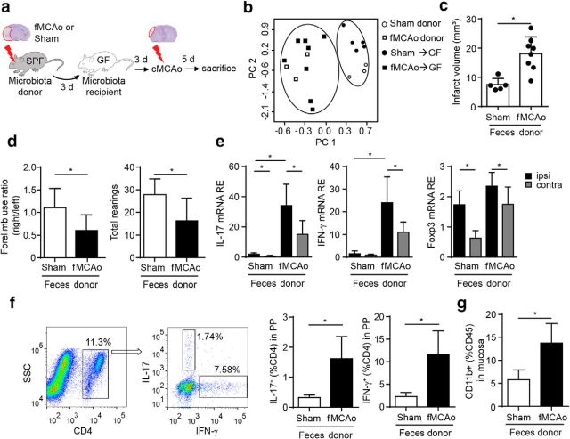
Brain ischemia-induced dysbiosis alters the poststroke immune reaction and exacerbates stroke outcome. a, Experimental design for recolonizing GF mice with gut microbiota from sham- or fMCAo- operated SPF donor mice. Three days after recolonization, mice underwent cMCAo induction and were killed another 5 d later for subsequent analysis. b, Principal component analysis of the microbiota composition in donor mice after sham and fMCAo surgery and microbiota composition in GF recipient mice 3 d after transplantation with donor microbiota. Analysis revealed a distinct pattern of the two donor populations before and after transplantation to recipient GF mice. c, Comparison of brain infarct volumes (cMCAo) in the recolonized recipient mice 5 d after cMCAo induction. d, Recolonizing mice with the gut microbiota obtained from post-fMCAo donors significantly increased brain lesion volumes and significantly reduced behavioral performance assessed by measuring forelimb use asymmetry (left) and total rearing activity (right) in the cylinder test (n = 5–8 per group, 2 individual experiments). e, Relative gene expression levels of IL-17, IFN-γ, and Foxp3 in the ipsilateral (ipsi) and contralateral (contra) hemispheres of GF recipient mice (n = 5–8 per group, 2 individual experiments). Recolonization with microbiota from fMCAo donor mice massively increased Th17 (IL-17) and Th1 (IFN-γ) expression compared with recipients of sham-microbiota. f, Representative dot plots with gating strategy for the flow cytometry analysis of Th17 (IL-17) and Th1 (IFN-γ) T cells in PPs (left). Percentages of IL-17+ and IFN-γ + T cells were significantly increased in PPs of mice recolonized with the fMCAo-microbiota (right panel). g, Percentage of CD11b+ monocytes/macrophages in the mucosal layer of the small intestine (terminal ileum) was significantly increased in mice receiving the microbiota from fMCAo donors compared with sham microbiota. All bar graphs: *p < 0.05, mean ± SD.

