Figure 5.
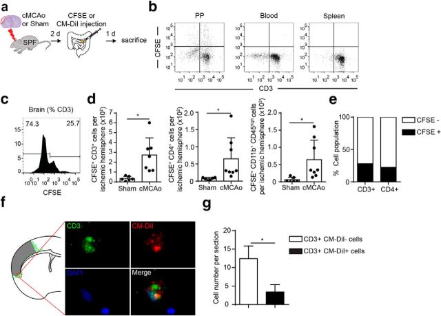
Lymphocytes migrate from PPs to the brain after stroke. a, Experimental design for tracing the migration of PP-derived lymphocytes in mice after cMCAo or sham surgery. CSFE or CM-DiI was injected in PP 2 d after the respective surgery and, 24 h later, brain and lymphoid organs were dissected and analyzed for dye-positive T cells. b, Validation of site-specific T-cell labeling with CFSE in PP 3 h after microinjection; T-cell labeling was not detectable in blood and spleen. c, Representative histogram for CFSE+ T cells (gated for CD45+CD3+ expression) from flow cytometry analysis of brain homogenates of the ipsilateral hemisphere 24 h after CFSE microinjection in all detectable PPs. d, Quantification of flow cytometry analysis shows increased numbers of CFSE+ total T cells (CD3+) and Thelper cells (CD4+) and monocytes (CD11b+) in ischemic hemispheres 3 d after cMCAo compared with sham control. e, Percentage of CFSE-labeled CD3+ and CD4+ T cells identified in ischemic hemispheres 3 d after cMCAo and 24 h after PP labeling. f, Brain-invading CM-DiI+ T cells derived from PPs were identified in the peri-infarct region and are illustrated as a cumulative map from five mice on one topographical coronal brain section at the bregma level g, Quantification of CM-DiI- and CM-DiI+ T cells (CD3+) per one histological brain section used to generate the cumulative map shown in f.

