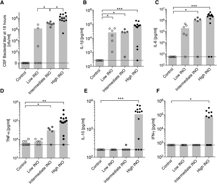
Figure 1.
Dose-dependent bacterial growth and cytokine profile in the CSF 18 h after infection with S. pneumoniae. A, Dose-dependent growth of S. pneumoniae in the CSF determined at 18 h after infection. B–F, The levels of IL-1β (B), IL-6 (C), TNF-α (D), IL-10 (E), and IFN-γ (F) were measured in the CSF 18 h after infecting the animals intracisternally with three different loads of S. pneumoniae (n = 5–13 animals per group). The means are indicated in the bar graphs. The dashed lines indicate the detection limit for each cytokine. An unpaired t test with Welch's correction was used for single comparisons. *p < 0.05; **p < 0.01; ***p < 0.001.

