Figure 6.
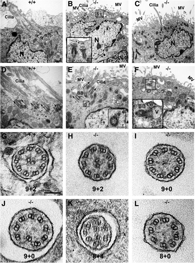
Ulk4 deficiency impairs axonemal structure and ciliary function. A–F, Transmission electron microscopic images of radial sections of ependymal cells. Clusters of multicilia with the same orientation appeared on P18 WT ependymal cells (A, D, arrows), but reduced (B, C, E) or absent (F) in Ulk4tm1a/tm1a cells (B, C, E, F). The black boxes in B and F show an immature cilium, namely a BB-vesicle fusion complex, beneath the Ulk4tm1a/tm1a cell membrane. Cilium length was not shortened in the Ulk4tm1a/tm1a mice (A, C–E), but the BB orientation was dispersed in the Ulk4tm1a/tm1a cells (A, D). Arrows show normally embedded BBs in WT (D), but differently oriented in the Ulk4tm1a/tm1a cells (E). The black box in E shows cross-sectional view of a BB foot in a vertical orientation. The black box in F shows a BB–vesicle fusion complex oriented parallel to the ependymal membrane, which varies by 90° from the normal orientation. G, WT littermates presented normal axonemes of 9 + 2. H–L, Some Ulk4tm1a/tm1a motile cilia exhibited 9 + 2 (H), but others either lost the central doublets without (9 + 0; I) or with disorganization of a peripheral doublet (also 9 + 0; J), or missed a peripheral doublet but had a supernumerary central doublet (8 + 4; K), or lost peripheral and central doublets (8 + 0; L). Scale bars: A–C, 1000 nm; D–F, 500 nm; G–L, 100 nm. Insets in B, E, and F were magnified three times from the respective images. MV, Microvilli; N, nucleus; V, vesicle; +/+, Ulk4+/+; −/−, Ulk4tm1a/tm1a.

