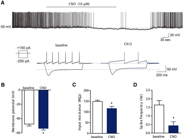
Figure 5.
Inhibition of hM4D-expressing VTA dopamine neurons following CNO application. A, Sample traces of spontaneous (top) and evoked action potentials (bottom) in hM4D-expressing dopamine neurons before and after CNO (10 μm) application. B–D, Group data for the effect of CNO on membrane potential (B), input resistance (C), and firing rate of the spontaneous action potentials (D; n = 7) in hM4D-expressing VTA dopamine neurons. Data represent mean ± SEM. *, Significant difference compared with the baseline.

