Figure 7.
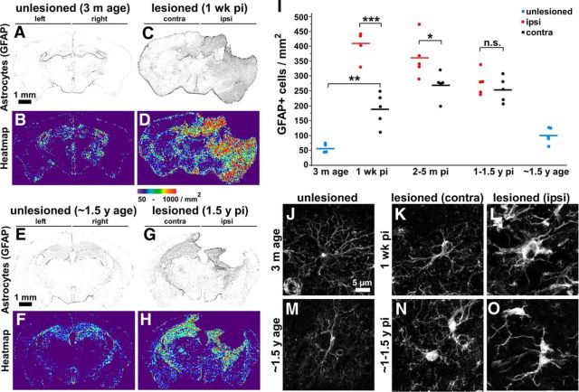
Increased astrocyte density in widespread brain regions in acute and chronic stages of TBI. GFAP+ astrocyte density and distribution were assessed in coronal brain sections. A–H, Representative images in grayscale and heat map: unlesioned young mice (A, B), mice at 1 week after CCI TBI (C, D), unlesioned old mice (E, F) and at 1.5 years after CCI TBI (G, H). I, Quantification of GFAP+ astrocyte density (average of entire hemibrain) from mice that were unlesioned or at indicated time points after CCI injury. Each dot represents one animal; n = 4, 5, 5, 5, 5, 5, 5, and 4 mice, respectively; mean values are indicated by horizontal lines. ***p < 0.001, *p < 0.05 calculated by one-way ANOVA test with Tukey–Kramer post hoc test between indicated groups. J–O, High-magnification confocal images of the astrocytes: unlesioned young mice (3 months age) (J), mice at 1 week after CCI TBI, contralateral (K) or ipsilateral (L), unlesioned old mice (∼1.5 years age) (M), and mice at 1.5 years after CCI TBI, contralateral (N) or ipsilateral (O). Note that astrocytes in unlesioned young mice have a star-shaped morphology with long processes (J). Acutely after injury, cell bodies of astrocytes become enlarged (L). In chronic stages (>1 year after injury), most of the astrocytes preserve their increased volume (N, O) compared with unlesioned age-matched mice (M).

