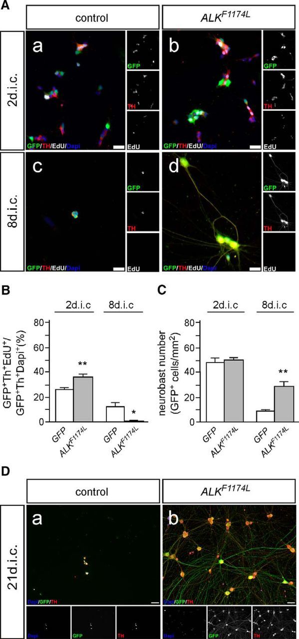
Figure 5.

Effects of activated ALK on cultured chick sympathetic neuroblasts. A, E7 sympathetic ganglion cells were transfected with expression vectors for GFP (control; Aa, Ac) or GFP and ALKF1174 L (Ab, Ad) and analyzed by GFP/Th/EdU-triple immunostaining after 2 (Aa, Ab) and 8 dic (Ac, Ad) for proliferating EdU-incorporating neuroblasts. Note the neuronal morphology induced by activated ALK signaling at 8 dic (Ad). B, Quantification reveals a significantly increased proliferation of ALKF1174L-expressing neuroblasts compared with GFP controls at 2 dic (mean ± SEM; n ≥ 6; **p < 0.01). At 8 dic, in contrast, proliferating neuroblasts are absent (mean ± SEM; n ≥ 6; *p < 0.05; unpaired two-tailed t test). C, ALKF1174L expression does not affect neuroblast number at 2 dic but results in a strongly increased survival of Alk-induced neurons (mean ± SEM; n ≥ 4; **p < 0.01). D, Sympathetic neurons induced by activated ALK survive for extended culture periods (3 weeks; Db), in contrast to GFP-transfected control neuroblasts (Da). Please note the large neuron cell bodies and extensive neurite network of ALK-induced sympathetic neurons. Scale bar, 50 μm.
