Figure 7.

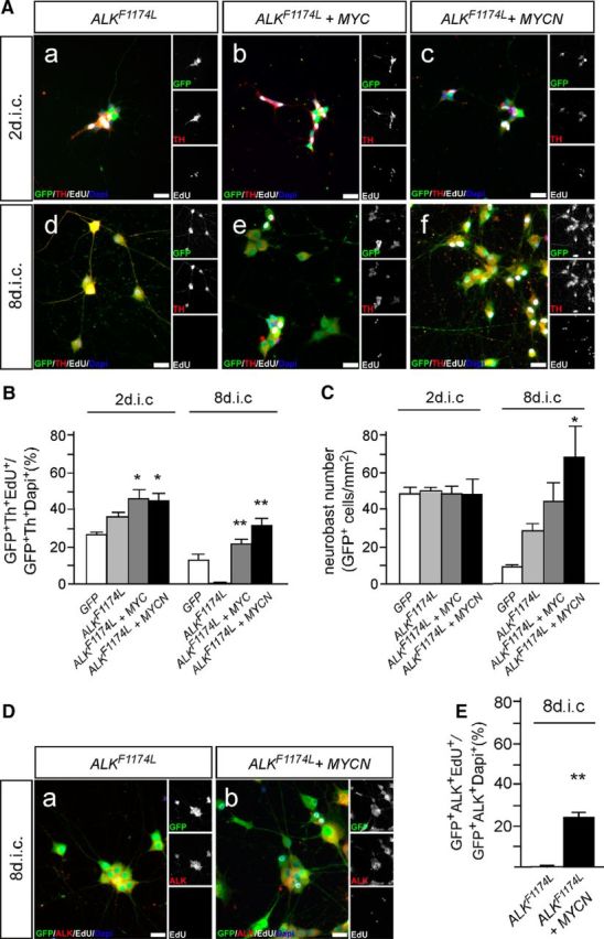
Coexpression of ALKF1174L and MYC proteins supports neuroblast proliferation and survival. A, E7 sympathetic ganglion cells were transfected with expression vectors for GFP and ALKF1174L (Aa, Ad); or GFP, ALKF1174L, and MYC (Ab, Ae); or GFP, ALKF1174L, and MYCN (Ac, Af); and analyzed by GFP/Th/EdU-triple immunostaining after 2 (Aa–Ac) and 8 dic (Ad–Af) for proliferating EdU-incorporating neuroblasts. Note that proliferating ALK/MYC cells (Ae, Af) are smaller and show a less mature neuronal morphology compared with neurons induced by activated ALK signaling at 8 dic (Ad). B, Quantification reveals a significantly increased proliferation of MYC/ALKF1174L-expressing and MYC/ALKF1174L-expressing neuroblasts compared with ALKF1174L neuroblasts at 2 dic (mean ± SEM; n ≥ 6; *p < 0.05). At 8 dic, proliferating neuroblasts are present in transfected cultures but absent in ALKF1174L neuroblasts (mean ± SEM; n ≥ 6; **p < 0.01; unpaired two-tailed t test). C, The combined MYCN/ALKF1174L expression does not affect neuroblast numbers at 2 dic, but results in a strongly increased neuron number at 8 dic compared with ALKF1174L (mean ± SEM; n ≥ 4; *p < 0.05). Data for GFP-transfected controls are included for comparison from Figure 5B,C. D, Proliferation of sympathetic neuroblasts transfected with expression vectors for GFP and ALKF1174L (Da); or for GFP, ALKF1174L, and MYCN (Db); and analyzed by GFP/ALK/EdU-triple immunostaining after 8 dic for proliferating EdU-incorporating ALK+ neuroblasts. E, Quantification reveals increased proliferation of ALK+ neuroblasts in MYCN/ALKF1174L compared with ALKF1174L cultures (mean ± SEM; n ≥ 3; **p < 0.01).
