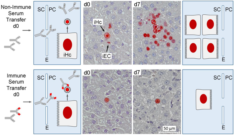
Fig. 3.
Virus-specific antibodies prevent cell-to-cell spread within host tissue. (Far left and far right panels) Explanatory charts of liver tissue compartments. SC sinusoidal compartment. E liver sinusoidal endothelium. PC parenchymal compartment. iHc infected hepatocyte. Unspecific and virus-specific (red-marked variable region) antibodies present in transferred sera enter liver parenchyma via fenestrae. Only virus-specific antibodies intercept released virions (Center images) Immunohistological images of liver tissue sections taken on day 0 (d0) and day 7 (d7) after serum transfer. iEC infected endothelial cell, a verified cellular site of MCMV latency [48]. Infected cells were identified by red staining of intranuclear IE1 protein. Conceptionally based on Wirtz et al. 2008, MMIM 197:151-158, with new tissue sections from stored embedded organs used for the original publication [47].

