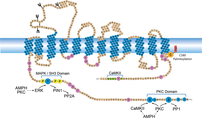
Figure 1. Phosphorylation characteristics of DAT.
Schematic diagram of rDAT showing PKC and MAPK domain phosphorylation sites in blue, with Ser7, Ser13, and Thr53 highlighted with large circles. Other intracellular Ser and Thr residues are shown in mauve, prolines flanking Thr53 that constitute an SH3 binding domain are shown in yellow, the CAMKII binding domain in the C-terminus is shown in green, and palmitoylation site Cys580 is shown in orange. Known and suspected kinase, phosphatase, and PIN1 inputs into Ser7, Ser13, and Thr53 are indicated with arrows, and dashed lines show indirect AMPH and PKC inputs into each site.

