
Figure 4.
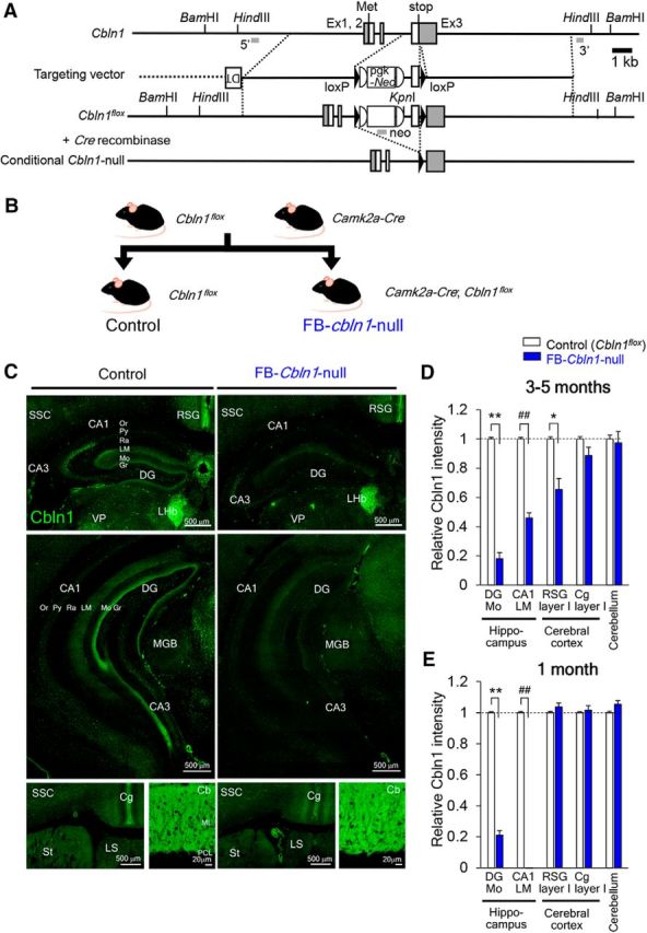
Generation and characterization of FB conditional Cbln1-null mice. A, Schematic representation of the Cbln1 genomic structure, targeting vector and targeted genes (Cbln1flox and conditional Cbln1-null). Filled triangles indicate loxP sequences. B, Breeding scheme. FB Cbln1-null (FB-Cbln1-null) mice were produced by crossing Cbln1flox/flox with Camk2a-Cre mice. C, Immunohistochemical analyses of Cbln1 expression in FB-Cbln1-null mice at 3–5 months of age. D, E, Cbln1 expression levels relative to those in control (Cbln1flox/flox) mice. At 3–5 months, Cbln1 expression in the hippocampus (DG-Mo and LM) layer I of RSG was reduced in FB-Cbln1-null mice (D). **p = 1.41 × 10⁻⁶; ##p = 0.0000338; *p = 0.0158. At 1 month, Cbln1 expression was also reduced in the hippocampus, but not in RSG (E). **p = 9.19 × 10⁻⁶; ##p = 0.0000949; n = 16 images for each from 3–4 animals, Mann–Whitney U test. Cbln1 expression in other brain regions, including Cg and cerebellum, was largely unaffected. For abbreviations, see the legend to Figure 3. Scale bars, 20 μm for the cerebellum and 500 μm for the rest of the panels.