Figure 5.
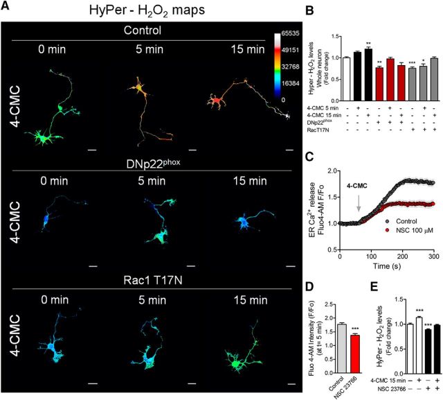
RyR stimulation induces H2O2 production through the NOX complex by a Rac1-dependent mechanism. Neurons were transfected after 1 DIV with the HyPer biosensor alone (control) or cotransfected with the DNp22phox or Rac1T17N (dominant-negative version of Rac1) constructs. After 1 d of expression, neurons were treated with 4-CMC. A, H2O2 maps after 750 μm 4-CMC stimulation for 5 and 15 min in control, DNp22phox-, and Rac1T17N-transfected neurons. B, Quantification of the HyPer-H2O2 levels of A. *p < 0.05, **p < 0.01, ***p < 0.001 versus control (white column), ANOVA, Dunnett's post test (15 neurons were analyzed for each condition). C, RyR-mediated Ca2+ release in control and NSC 23766-treated neurons (100 μm). D, Quantification of the F/F0 ratio at t = 5 min of C. ***p < 0.001 versus control, Student's t test (a total of 60 neurons were analyzed). E, Neurons (2 DIV) expressing the HyPer biosensor were treated with NSC 23766 (100 μm) for 1 h and then stimulated with 4-CMC for 15 min to measure HyPer-H2O2 levels. ***p < 0.001 versus control, ANOVA, Dunnett's post test. Results are from 3 different independent cultures (n = 3). Scale bar, 20 μm.

