Figure 1.
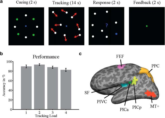
Design of the attentional tracking task. a, Each tracking trial started with a cueing phase (2 s), during which eight disks were arranged radially in the visual periphery and remained stationary. A subset of disks (either 1, 2, 3, or 4) was temporarily highlighted in green and served as the tracked targets (in the current example, 4 targets had to be tracked). The more targets that were tracked, the greater was the attentional load. After brief highlighting, the targets turned white, and all disks moved randomly, at a constant speed, across the screen. Disks never overlapped or collided and were repelled from each other and from the borders of the display. Participants were instructed to maintain central fixation and to track the targets covertly, using their attention. After 14 s of tracking, all disks stopped moving, and one disk was highlighted in blue. Participants were given 2 s to indicate whether the blue disk was a tracked target or a distractor. At the end of each trial, feedback (green or red fixation for correct or incorrect responses) was provided for 2 s. In the primary experiment, each tracking trial was followed by a baseline trial, during which the disks moved randomly across the screen for 20 s, while participants maintained central fixation. On baseline trials, none of the disks was highlighted and participants passively viewed the moving disks. In the control experiment, each tracking trial was followed by a blank baseline, during which participants maintained central fixation. b, Average behavioral performance (with SE) in each of the four tracking conditions of the primary experiment (25 participants; chance level, 50%; see Materials and Methods for details). c, Locations of ROIs, shown on the inflated left hemisphere of a typical participant (dark gray, sulci; light gray, gyri). SF, Sylvian fissure (also called lateral sulcus); PICa/PICp, anterior/posterior clusters of the PIC area; PPC, posterior parietal cortex; MT+ is V5. The PIVC was defined by means of caloric stimulation, whereas the PIC and MT+ were defined based on stimulation with visual motion. The FEF was localized by means of activation during saccades. The PPC was defined based on anatomical landmarks (parietal gyrus and sulcus).

