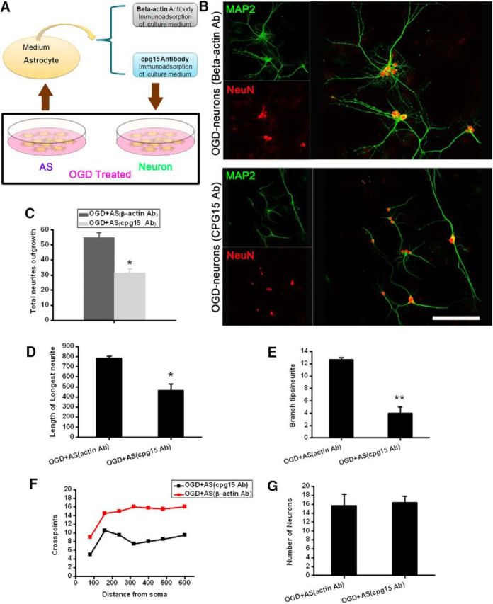
Figure 4.

Immunoadsorption of cpg15 from the culture medium of OGD-treated hippocampal astrocytes suppresses dendritic outgrowth of OGD-injured primary hippocampal neurons and reestablishment of neural network. A, Schematic diagram showing the immunoadsorptional removal of soluble cpg15 from OGD-treated astrocyte culture medium. B, Representative immunofluorescence double staining of dendritic marker MAP2 (green) and neuronal marker NeuN (red) at reoxygenation 48 h in 4 h OGD-injured primary cultured neurons cultured with the cpg15-immunoadsorbed medium of OGD-treated astrocytes (cpg15 Ab). β-Actin immunoadsorbed medium as the control (β-actin Ab). C–G, Statistical data of total neurite density (C), length of longest neurites (D), branch tips per neurite (E), crosspoints (F), and number of total neurons (G) from B. Scale bars, 50 μm. n = 10 for each group. *p < 0.05. **p < 0.01.
