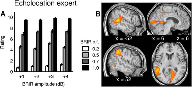
Figure 8.
Blind echolocation expert. A, Psychophysical performance as in Figure 5A. Bars represent the mean room size classification as a function of BRIR compression factor (grayscale of the bars) and BRIR amplitude (bar groups). Error bars indicate SEs across trial repetitions. Without any prior training, the classification is very stable and similar to that of the extensively trained, sighted subjects. B, Regions of activity in an echolocation expert during active echolocation compared with sound production without auditory feedback. The strongest regions of activations in the fMRI data were found in visual and parietal areas (compare Table 4). Activity maps were thresholded at p < 0.05 (FDR corrected) and overlaid on the subject's normalized structural image.

